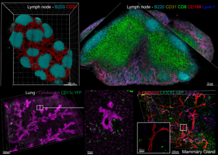
The Center for Advanced Tissue Imaging (CAT-I) is an NIAID and NCI supported effort involving collaborative studies between experts in the Laboratory of Immune System Biology, DIR, NIAID and investigators in the DIR, NIAID and CCR, NCI.
Read more about the Center for Advanced Tissue Imaging (CAT-I).
Major Areas of Focus
- To enable collaboration in the application of LBS methods of Histo-cytometry in 2D and 3D
- To enable highly multiplex, quantitative image analysis of tissues samples, primarily but not exclusively of human origin
- To advance our understanding of immune processes in various diseases
- To develop better vaccines
Work With Us
CAT-I is intended to support the application of evolving advanced imaging methods to multiplex, high-dimension tissue analysis. The complexity of such research requires that the projects be undertaken as collaborations and not ‘fee for service’ activities. This will in turn influence how projects are selected for CAT-I participation.
- Read more about the criteria we use to select projects
- Propose a project using our collaboration proposal form (The third round of proposals will be accepted through March 4, 2019, with decisions expected within a month of the closing date. Proposals should be sent to rgermain@niaid.nih.gov with the subject line “CAT-I proposal submission.”)
- Read more about CAT-I operating principles before you consider submitting a proposal